CHANNELS


Dr. med. Matthias J. Hackl
+49 221 478-32319
matthias.hackl@uk-koeln.de
TEAM MEMBERS
Julia Binz-Lotter, PhD
Agnes Hackl, MD/PhD
Nelli Rutkowski, M.Sc.
Matthias Hackl
In vivo imaging of the mouse glomerulus
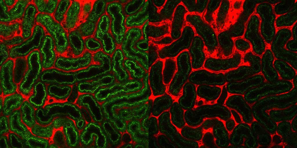
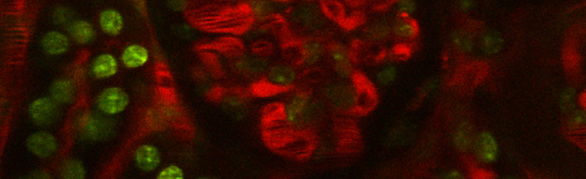
Glomerular filtration is dependent on the complex interplay of different cell types, including glomerular endothelial cells, mesangial cells, and podocytes. The interactions of these cells facing high blood flow and filtration pressure can not be adequately studied in in vitro model systems. We therefore use multiphoton microscopy to visualize glomerular processes in living animals. Mice with genetically encoded calcium indicators enable us to study changes in intracellular calcium levels of glomerular cells in health and disease. Using this technique, we were able to show that healthy podocytes rarely react to Angiotensin II with a calcium signal. However, after the induction of disease podocytes are much more susceptible to Angiotensin II-mediated calcium signals. This underlines the importance of the inhibition of the Renin-Angiotensin-Aldosterone-System using ACE inhibitors or Angiotensin receptor blockers in patients with kidney disease.
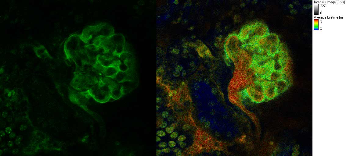
While the biological function of cGMP pathways is well described in vascular smooth muscle cells, very little is known about their role in the kidney. To study the role of cGMP signaling in podocytes and glomerular endothelial cells and investigate the crosstalk between these cells, we employ mouse lines with FRET-based cGMP indicators allowing imaging approaches based on ratiometric fluorescence intensity and FLIM-FRET.
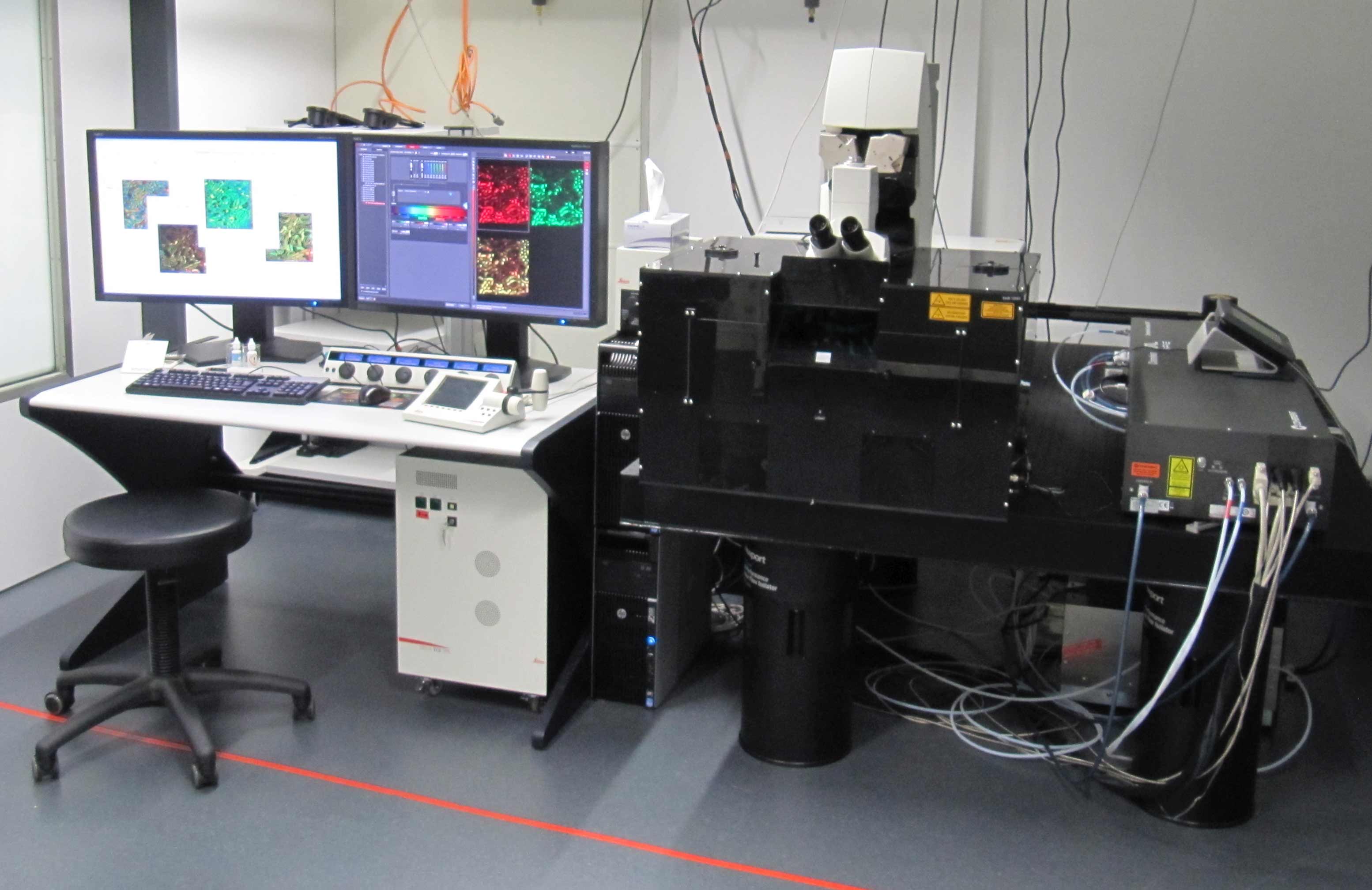
For our experiments, we use state-of-the-art equipment of the CECAD Imaging Facility, including a Leica TCS SP8 multiphoton microscope. Together with our innovative mouse models, this enables us to obtain unprecedented views of glomerular biology.
SELECTED PUBLICATIONS
Butt, L., Unnersjo-Jess, D., Hohne, M., Edwards, A., Binz-Lotter, J., Reilly, D., Hahnfeldt, R., Ziegler, V., Fremter, K., Rinschen, M.M., Helmstadter, M., Ebert, L.K., Castrop, H., Hackl, M.J., Walz, G., Brinkkoetter, P.T., Liebau, M.C., Tory, K., Hoyer, P.F., Beck, B.B., Brismar, H., Blom, H., Schermer, B., and Benzing, T. (2020) A molecular mechanism explaining albuminuria in kidney disease. Nat Metab, 2(5): 461-474.
Binz-Lotter, J., Jungst, C., Rinschen, M.M., Koehler, S., Zentis, P., Schauss, A., Schermer, B., Benzing, T., and Hackl, M.J. (2020) Injured Podocytes Are Sensitized to Angiotensin II-Induced Calcium Signaling. J Am Soc Nephrol, 31(3): 532-542.
Koehler, S., Brahler, S., Kuczkowski, A., Binz, J., Hackl, M.J., Hagmann, H., Hohne, M., Vogt, M.C., Wunderlich, C.M., Wunderlich, F.T., Schweda, F., Schermer, B., Benzing, T., and Brinkkoetter, P.T. (2016) Single and Transient Ca(2+) Peaks in Podocytes do not induce Changes in Glomerular Filtration and Perfusion. Sci Rep, 6: 35400.
Burford, J.L., Villanueva, K., Lam, L., Riquier-Brison, A., Hackl, M.J., Pippin, J., Shankland, S.J., and Peti-Peterdi, J. (2014) Intravital imaging of podocyte calcium in glomerular injury and disease. J Clin Invest, 124(5): 2050-8.
Hackl, M.J., Burford, J.L., Villanueva, K., Lam, L., Susztak, K., Schermer, B., Benzing, T., and Peti-Peterdi, J. (2013) Tracking the fate of glomerular epithelial cells in vivo using serial multiphoton imaging in new mouse models with fluorescent lineage tags. Nat Med, 19(12): 1661-6.